Attention: Restrictions on use of AUA, AUAER, and UCF content in third party applications, including artificial intelligence technologies, such as large language models and generative AI.
You are prohibited from using or uploading content you accessed through this website into external applications, bots, software, or websites, including those using artificial intelligence technologies and infrastructure, including deep learning, machine learning and large language models and generative AI.
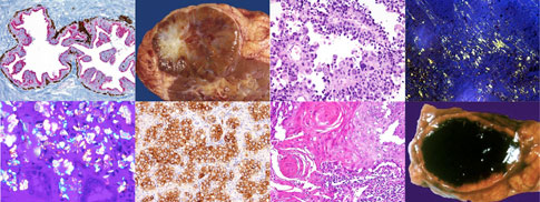
Pathology for Urologists
This program was designed to help Urology residents and fellows familiarize themselves with the pathologic features of common urologic entities. This will serve not only as a resource tool for your review but also as a quick reference guide to urologic pathology.
This tutorial covers >250 different entities, encompassing pertinent histoanatomic structures to recent innovations and advances in the field. These include among others, newly recognized tumors and terminologies, latest classification schemes, current grading approaches (e.g. recent WHO grading for urothelial neoplasms, ISUP modified Gleason grading, etc.), molecular alterations, and commonly used ancillary diagnostic techniques particularly immunohistochemistry. Main differential diagnoses and their distinguishing features are also presented. There are >650 high-quality images which include gross pictures, histologies, cytologies, special stains, other ancillaries, drawings and illustrations. A self-test is provided at the end for your own assessment.
Descriptions are made short and concise (not >1 page per entity) but enough to cover the basics that urologists should know about pathology. The text is bulleted, key terms and messages are bolded or italicized, and some pathology lexicons are clarified. The images have labels in place and can be enlarged for ease of use in your laptops, tablets and even smartphones.
I like to acknowledge Dr. Jo Ellen Cottey Krueger who wrote the previous version together with Dr. Herb Hagler, Dr. Key Stage and Dr. Warren Snodgrass. A significant amount of their original contributions including their fantastic images and drawings are retained in this updated version reflective of the excellent work they had accomplished.
I also would like to thank my residents and fellows in both Urology and Pathology for providing me with the inspiration to accomplish this task.
Click on the table of contents to select the entities. Enjoy!
Gladell P. Paner, MD
Pathology and Section of Urology, Surgery
University of Chicago
August 2012
advertisement
advertisement