Attention: Restrictions on use of AUA, AUAER, and UCF content in third party applications, including artificial intelligence technologies, such as large language models and generative AI.
You are prohibited from using or uploading content you accessed through this website into external applications, bots, software, or websites, including those using artificial intelligence technologies and infrastructure, including deep learning, machine learning and large language models and generative AI.
Neurofibroma
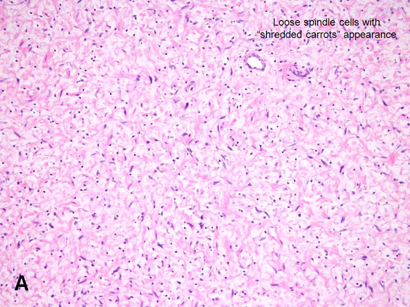
Image A
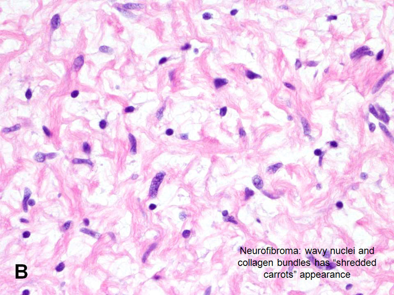
Image B
- Extremely rare, occur in patients with neurofibromatosis I.
- GU is involved in ~20% in patients with neurofibromatosis.
- Bladder is most commonly involved in GU tract, and involvement can be extensive.
- Typically involves young patients (~1/3 pediatric patients); male>female (3:1).
- Histology:
- Immunohistochemistry: S100+.
advertisement
advertisement